Patellar Instability (Unstable Kneecap)
General Information
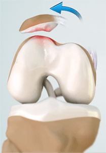
The patella, also known as the “knee cap” sits at the front part of the knee. As the knee bends, it glides in a groove of the femur called the “trochlear groove”. This groove accommodates the patella so that it smoothly tracks in a straight line. When the patella slips out of this groove, the condition is called “patellar instability”.
Causes
In the normal knee, there is a deep trochlear groove in which the patella glides, allowing it to stay in position. In some people, however, the trochlear groove and the reciprocal shape of the patella can be abnormal.
This predisposes them to having the patella potentially coming out of place. The other reasons include having a weak quadriceps muscle, having lax joint or knocked knees. This condition may run in families. Some times, there are no underlying factors except a significant traumatic injury to your knee.
Symptoms & Diagnosis
The first symptom of patellar instability may be an injury where the patella slips to the outside part of the knee. This commonly occurs when the knee is in flexed position and the body is rotating. In its most severe circumstance, the person notes the patella sitting on the outside part of the femur.
An obvious lump is noted on the outer aspect of your leg and this is accompanied by severe pain. Sometimes the patella goes back into its position by itself as the knee is straightened. Not uncommonly you may need to go to hospital in order to have the patella put back into place (“re-located”).
A more subtle condition is when the patella partially slides out of the trochlear groove and then spontaneously goes back into position. This is known as “patellar subluxation”.
Symptoms of patellar instability include a feeling of buckling of the knee, a feeling of apprehension as you bend and twist especially at the front part of the knee, intermittent pain, swelling and clicking. Some people only have one episode of patellar instability. Occasionally it can become a recurrent problem.
Diagnosis
The diagnosis of patellar instability can be established by your doctor getting an accurate account of the events which led to your first episode of patellar instability. A detailed physical examination is important to establish the reason for your problem and to assess for other alignment issues and muscle weakness. Your doctor will assess how the patella tracks in the trochlear groove of the femur, whether there are tight ligaments on the outside of the knee contributing the patella instability or whether there is weakness of the inner quadriceps muscle (VMO) which can prevent this condition from happening.
Investigations are usually required. This includes a series of x-rays to look at the shape of the patella and trochlear groove. Sometimes an MRI (magnetic resonance imaging) scan is ordered to assess the cartilage lining of the joint to ensure that this has not been damaged and to assess the soft tissues which hold the patella in place.
Treatment
After your first episode of patellar instability it is important to institute the basic principles of soft tissue management – RICE: Rest, Ice, Compression, Elevation. Once your pain and swelling subsides, it is important to start exercises and often the best place to start is a rehabilitation program with a physiotherapist.
The aims are to reduce swelling, re-establish joint motion followed by a strengthening program of the quadriceps muscle, especially its main inner portion, the vastus medialis obliquus (VMO) muscle. You also may be recommended stretching exercises of your hamstring muscles and tight ligaments on the outer part of your knee. Occasionally taping of the patella into place may help relieve or reduce the chance of ongoing symptoms.
Exercise bike work and a gym based program focusing on leg press and mini squats will aid in re-establishing joint muscle strength. Occasionally a stabilising brace may be needed as well.
When is surgery required?
If patellar instability becomes a recurrent problem, corrective surgery may be recommended. This often requires a reconstruction of a ligament on the inner part of the patella called the “medial patellofemoral ligament”.
This ligament helps hold the patella in the trochlear groove. One of your hamstring tendons (autograft) is used to do this. In conjunction with this, a “lateral release” may be required. This relieves tight structures on the outer aspect of the patella and helps patellar tracking in some patients.
If there are severe abnormalities in tracking of the patella in the trochlear groove, your surgeon may also recommend a procedure called a “tibial tubercle osteotomy”. This involves moving a segment of bone at the lower front part of the knee where the patella tendon inserts. The aim is to allow the patella to track in a more central position. It may be combined with the two other procedures mentioned above.