Medial Collateral Ligament (MCL) Tears
General Information
What are the collateral ligaments?
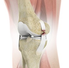
The collateral ligaments are structures that provide side-to-side stability to the knee joint. Unlike the cruciate ligaments which are inside the knee joint, the collateral ligaments lie outside of the joint.
The medial collateral ligament (MCL) connects the inner aspect of the thigh bone (femur) to the inner aspect of the shin bone (tibia) and provides stability to the inner side of the knee. The lateral collateral ligament (LCL) connects the outer aspect of the femur to the fibula bone and stabilises the outer aspect of the knee.
How does the MCL get injured?
Injuries to the MCL are usually caused by contact on the outer side of the knee and are accompanied by sharp pain on the inside of the knee. The patient’s knee commonly buckles inwards, stretching the MCL or completely tearing it. The LCL is rarely injured.
The MCL is assessed by your doctor or physiotherapist by applying a stress to the tibia bone and seeing if the leg turns outwards. This often reproduces pain but also instability of the knee, especially when compared to the opposite normal knee.
Symptoms & Diagnosis
What symptoms might I experience from an MCL tear?
Patient’s with MCL tears usually experience sudden pain on the inner aspect of the knee. This may be associated with a tearing or popping sensation at the time of injury. Swelling and bruising usually accompany these injuries. Sometimes placing weight on the leg is initially painful.
Diagnosis
Often these tears can be diagnosed clinically, without requiring any further tests. If the diagnosis is uncertain or other conditions need to be excluded, your doctor may order x-rays and an MRI scan to confirm the diagnosis.